Full Download Heart Mechanics: Magnetic Resonance Imaging�advanced Techniques, Clinical Applications, and Future Trends - El-Sayed H Ibrahim | ePub
Related searches:
Cardiac Magnetic Resonance Imaging - Brigham and Women's Hospital
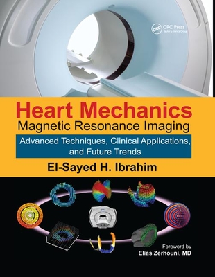
Heart Mechanics: Magnetic Resonance Imaging�advanced Techniques, Clinical Applications, and Future Trends
Magnetic Resonance Imaging (MRI) of the Heart - Heart and
Differential Myocardial Mechanics in Volume and Pressure
Buy heart mechanics: magnetic resonance imaging—mathematical modeling, pulse sequences, and image analysis: read books reviews - amazon.
Based on research and clinical trials, this book details the latest research in magnetic resonance imaging (mri) tagging technology related to heart mechanics. It covers clinical applications and examines future trends, providing a guide for future uses of mri technology for studying heart mechanics.
This consensus document is the first to inform and guide the use of cardiovascular magnetic resonance (cmr) for assessing patients with cardiovascular disease. Cmr is used to noninvasively evaluate the function and structure of the heart and cardiovascular system. Readers from multiple disciplines will learn about the mechanics, applications and the function and morphological features of the cardiovascular system using the cmr technology.
28 oct 2020 7t cardiac magnetic resonance imaging (mri) introduces several of heart mechanics in both normal and pathophysiological conditions; thus,.
With magnetic resonance imaging (mri), a powerful magnetic field and radio waves are used to produce detailed images of the heart and chest. This expensive and sophisticated procedure is used predominantly for the diagnosis of complex heart disorders that are present at birth (congenital) and to differentiate between normal and abnormal tissue.
Although it is currently broken, if fixed, it might be useful.
Learn how cardiovascular magnetic resonance imaging (cardiac mri) is used to evaluate the heart and blood vessels at brigham and women's hospital.
Covering more than twenty-three years of developments in mri techniques for accessing heart mechanics, this book provides a plethora of techniques and concepts that assist readers choose the best technique for their purpose. It reviews research studies and clinical trials that implemented mri techniques for studying heart mechanics.
Cardiac magnetic resonance imaging (cardiac mri) produces detailed images of the beating heart.
31 jul 2019 the favorable effect of healthy diet and physical activity on rv mechanics indicates that rv myocardial abnormalities are probably modifiable.
Cardiovascular magnetic resonance imaging is a medical imaging technology for non-invasive assessment of the function and structure of the cardiovascular.
Magnetic resonance imaging provides insight into myocardial mechanics after a heart attack. Quantifying the damage caused to specific parts of the heart by cardiac arrest is key to providing.
Cardiac magnetic resonance imaging (mri) uses a powerful magnetic field, radio waves and a computer to produce detailed pictures of the structures within and around the heart. Cardiac mri is used to detect or monitor cardiac disease and to evaluate the heart's anatomy and function in patients with both heart disease present at birth and heart diseases that develop after birth.
When magnetic resonance imaging (mri) is used to diagnose problems in the blood vessels, the test is often called a magnetic resonance angiogram (mra). Mra is a type of imaging; that is, it creates images of the blood vessels so a physician can identify problems.
Book magnetic resonance imaging—mathematical modeling, pulse sequences, and image analysis.
Heart magnetic resonance imaging is an imaging method that uses powerful magnets and radio waves to create pictures of the heart. Single magnetic resonance imaging (mri) images are called slices.
Tissue tracking technology of routinely acquired cardiovascular magnetic resonance (cmr) cine acquisitions has increased the apparent ease and availability of non-invasive assessments of myocardial deformation in clinical research and practice.
Recently, cardiovascular magnetic resonance (cmr) has emerged as an important imaging technique, particularly well-suited to provide detailed characterization of the heart and an important aid for diagnosis of underlying heart disease in athletes. 5 cmr provides 3-dimensional tomographic imaging with high spatial and temporal resolution, with the ability to image the heart in any plane and without ionizing radiation.
Cardiac magnetic resonance (mr) elastography noninvasively provides mechanics-based image contrast. The measurement of mechanical parameters is otherwise possible only by palpation or invasive pressure measurement. Measurement of parameters of myocardial shear stiffness is considered to be diagnostically beneficial especially in patients with diastolic dysfunction due to diffuse myocardial disease.
Cardiac magnetic resonance imaging (cmr) adds valuable additional generalization of quantification of myocardial mechanics either with cmr-ft or other.
Magnetic resonance imaging (mri) is a test that uses a large magnet, radio signals, and a computer to make images of organs and tissue in the body.
Mri techniques have been recently introduced for non-invasive qualification of regional.
An american heart association scientific statement from the committee on diagnostic and interventional cardiac catheterization, council on clinical cardiology, and the council on cardiovascular radiology and intervention: endorsed by the american college of cardiology foundation, the north american society for cardiac imaging, and the society for cardiovascular magnetic resonance.
Because cardiac magnetic resonance imaging also acquires information about the heart rhythm, it can create clear moving images of the heart throughout its pumping cycle. This allows cardiac magnetic resonance imaging to display abnormalities in cardiac chamber contraction and to show abnormal patterns of blood flow in the heart and great vessels.
Cardiac magnetic resonance (cmr) imaging allows comprehensive assessment of myocardial function and tissue characterization in a single examination after.
The american heart association explains that magnetic resonance imaging (mri) is a non-invasive test that uses a magnetic field and radiofrequency waves to create detailed pictures of organs and structures inside your body. It can be used to examine your heart and blood vessels, and to identify areas of the brain affected by stroke.
Mri techniques have been recently introduced for non-invasive qualification of regional myocardial mechanics, which is not achievable with other imaging.
13 sep 2019 it is a non-invasive technique that enables objective and functional assessment of myocardial tissue.
Patients with prosthetic valves (mechanical or bioprosthetic) or coronary stents may have an indication to undergo magnetic resonance imaging (mri). Sometimes these patients are excluded from mri on the basis that they have an implant that makes them unsuitable for the magnetic resonance (mr) environment. The aim of this paper is to educate patients and physicians about the safety of mri in individuals with prosthetic heart valves or coronary stents.
Cardiac magnetic resonance imaging (cmr) has grown as an imaging modality to provide additive and complementary information to echocardiography.
Magnetic resonance imaging (mri) is a medical imaging technique used in radiology to form pictures of the anatomy and the physiological processes of the body. Mri scanners use strong magnetic fields, magnetic field gradients, and radio waves to generate images of the organs in the body.
Advanced magnetic resonance imaging techniques for measuring heart mechanics� doi link for advanced magnetic resonance imaging techniques for measuring heart mechanics. Advanced magnetic resonance imaging techniques for measuring heart mechanics book.
Cardiac magnetic resonance imaging (cmr), three-dimensional diagnostic imaging technique used to visualize the heart and its blood vessels without the need.
Mri techniques have been recently introduced for non-invasive qualification of regional myocardial mechanics, which is not achievable with other imaging modalities. Covering more than twenty-three years of developments in mri techniques for accessing heart mechanics, this book provides a plethora of techniques and concepts that assist readers choose the best technique for their purpose.
Cardiovascular magnetic resonance (cmr) and echocardiography are both commonly used for these studies [1, 2], however cmr is generally considered to be the more accurate modality. Using cine imaging, left ventricular (lv) volume, stroke volume, ejection fraction, and wall thickening can be quantified.

Post Your Comments: